File:Electron_microscopy_reveals_mitochondrial_DNA_in_discrete_foci.jpg
From Wikipedia, the free encyclopedia
Size of this preview: 464 × 600 pixels. Other resolutions: 186 × 240 pixels | 371 × 480 pixels | 956 × 1,236 pixels.
Original file (956 × 1,236 pixels, file size: 248 KB, MIME type: image/jpeg)
| This is a file from the Wikimedia Commons. Information from its description page there is shown below. Commons is a freely licensed media file repository. You can help. |
| DescriptionElectron microscopy reveals mitochondrial DNA in discrete foci.jpg |
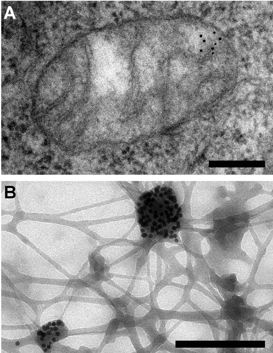
English: Electron microscopy reveals mitochondrial DNA in discrete foci. Bars: 200 nm. (A) Cytoplasmic section after immunogold labelling with anti-DNA; gold particles marking mtDNA are found near the mitochondrial membrane. (B) Whole mount view of cytoplasm after extraction with CSK buffer and immunogold labelling with anti-DNA; mtDNA (marked by gold particles) resists extraction. Iborra et al. BMC Biology 2004 2:9 doi:10.1186/1741-7007-2-9 |
| Date | |
| Source | The functional organization of mitochondrial genomes in human cells |
| Author | Francisco J Iborra1 , Hiroshi Kimura2 and Peter R Cook |
| Permission (Reusing this file) |
© 2004 Iborra et al; licensee BioMed Central Ltd. This is an Open Access article: verbatim copying and redistribution of this article are permitted in all media for any purpose, provided this notice is preserved along with the article's original URL. |
This file is licensed under the Creative Commons Attribution 2.0 Generic license.
- You are free:
- to share – to copy, distribute and transmit the work
- to remix – to adapt the work
- Under the following conditions:
- attribution – You must give appropriate credit, provide a link to the license, and indicate if changes were made. You may do so in any reasonable manner, but not in any way that suggests the licensor endorses you or your use.
Captions
Add a one-line explanation of what this file represents
Items portrayed in this file
depicts
2004
File history
Click on a date/time to view the file as it appeared at that time.
| Date/Time | Thumbnail | Dimensions | User | Comment | |
|---|---|---|---|---|---|
| current | 08:28, 26 January 2010 | | 956 × 1,236 (248 KB) | CopperKettle | {{Information |Description={{en|1=Electron microscopy reveals mitochondrial DNA in discrete foci. Bars: 200 nm. (A) Cytoplasmic section after immunogold labelling with anti-DNA; gold particles marking mtDNA are found near the mitochondrial membrane. (B) W |
File usage
The following pages on the English Wikipedia use this file (pages on other projects are not listed):
Global file usage
The following other wikis use this file:
- Usage on ar.wikipedia.org
- Usage on be.wikipedia.org
- Usage on bs.wikipedia.org
- Usage on el.wikipedia.org
- Usage on et.wikipedia.org
- Usage on eu.wikipedia.org
- Usage on fa.wikipedia.org
- Usage on fr.wikipedia.org
- Usage on gl.wikipedia.org
- Usage on id.wikipedia.org
- Usage on ja.wikipedia.org
- Usage on kk.wikipedia.org
- Usage on ko.wikipedia.org
- Usage on ru.wikipedia.org
- Usage on sl.wikipedia.org
- Usage on zh.wikipedia.org
Metadata
This file contains additional information, probably added from the digital camera or scanner used to create or digitize it.
If the file has been modified from its original state, some details may not fully reflect the modified file.
| _error | 0 |
|---|