File:Rostral_migratory_stream_mouse.jpg
From Wikipedia, the free encyclopedia
Size of this preview: 347 × 599 pixels. Other resolutions: 139 × 240 pixels | 278 × 480 pixels | 445 × 768 pixels | 1,196 × 2,063 pixels.
Original file (1,196 × 2,063 pixels, file size: 241 KB, MIME type: image/jpeg)
| This is a file from the Wikimedia Commons. Information from its description page there is shown below. Commons is a freely licensed media file repository. You can help. |
| DescriptionRostral migratory stream mouse.jpg |
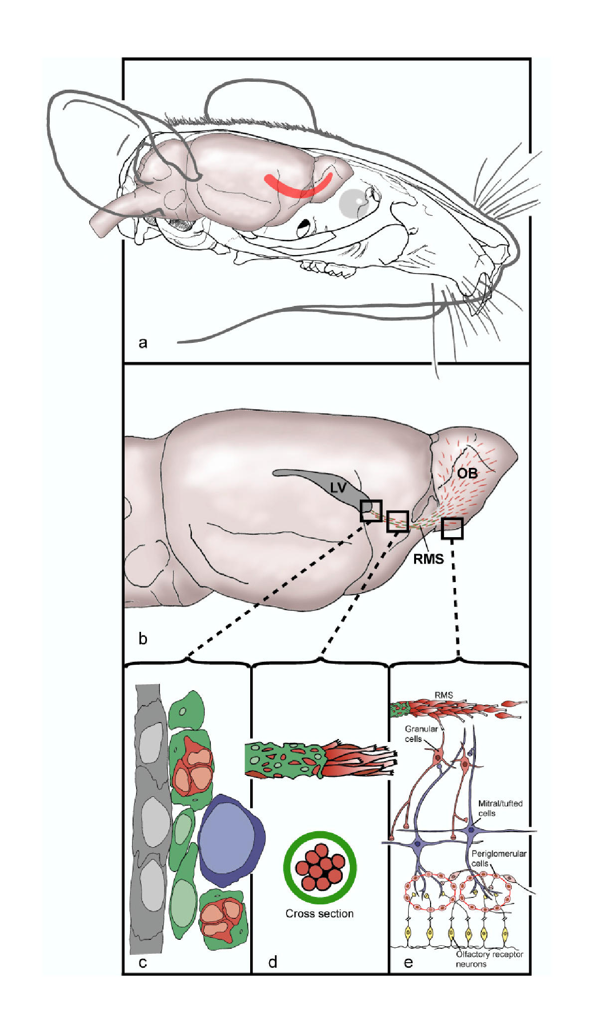
English: (a) Head of a mouse showing the location of the brain and the rostral migratory stream, RMS, (in red) along which newly generated neuroblasts migrate from the SVZ of the lateral ventricle into the olfactory bulb (OB). (b) The migration of newly generated neuroblasts begins at the lateral ventricle, continues along the RMS and terminated in the OB, where mature interneuron populations are generated. (c) Schematic based on electron microscopy showing the cytoarchitecture of the SVZ along the ventricle. Ependymal cells (gray) form a monolayer along the ventricle with astrocytes (green), neuroblasts (red) and transitory amplifying progenitors (TAP) (purple) comprising the SVZ. (d) Schematic showing the migration of neuroblasts along the RMS. Astrocytes (green) ensheath the migrating neuroblasts (red) and are thought to restrict and contain the neuroblasts to their specific pathway. (e) Migrating neuroblasts enter the OB, migrate radially and give rise to granule or periglomerular cells.
Русский: (A): Голова мыши. Показано расположение рострального миграционного тракта в мозге (красная полоса). По этому пути мигрируют свежесозданные нейробласты из субвентрикулярной зоны в обонятельную луковицу. (B): Миграция новых нейробластов начинается с бокового желудочка, затем клетки проходят весь РМТ до обонятельной луковицы, в которой генерируются взрослые популяции нейронов. (C): Схема, основанная на данных электронной микроскопии, демонстрирующая цироархитектуру субвентрикулярной зоны вдоль желудочка. Эпендимоциты (серые) формируют моно-слой а астроциты (зел), нейробласты (красн) и транзиторные делящиеся предшественники (TAP, пурпурные), составляют тело субвентрикулярной зоны. (D): Схема нейромиграции в тракте. Астроциты (зел.) окутывают мигрирующие нейробласты (красн.) и, как считается, ограничивают их свободу, направляя по строго предопределенному пути. (E): Мигрирующие нейробласты достигли обон. луковицы, теперь они мигрируют радиально и закрепляются, превращаясь в гранульные или перигломерулярные клетки. |
| Date | |
| Source | Lennington et al. Neural stem cells and the regulation of adult neurogenesis. Reproductive Biology and Endocrinology 2003 1:99 doi:10.1186/1477-7827-1-99 |
| Author | Jessica B Lennington, Zhengang Yang and Joanne C Conover |
| Permission (Reusing this file) |
© 2003 Lennington et al; licensee BioMed Central Ltd. This is an Open Access article: verbatim copying and redistribution of this article are permitted in all media for any purpose, provided this notice is preserved along with the article's original URL. |
| Other versions | Derivative works of this file: Rostral migratory stream mouse cropped.jpg |
This file is licensed under the Creative Commons Attribution 2.0 Generic license.
- You are free:
- to share – to copy, distribute and transmit the work
- to remix – to adapt the work
- Under the following conditions:
- attribution – You must give appropriate credit, provide a link to the license, and indicate if changes were made. You may do so in any reasonable manner, but not in any way that suggests the licensor endorses you or your use.
Captions
Add a one-line explanation of what this file represents
Items portrayed in this file
depicts
13 November 2003
image/jpeg
File history
Click on a date/time to view the file as it appeared at that time.
| Date/Time | Thumbnail | Dimensions | User | Comment | |
|---|---|---|---|---|---|
| current | 11:41, 4 August 2008 | | 1,196 × 2,063 (241 KB) | CopperKettle | {{Information |Description={{en|1=(a) Head of a mouse showing the location of the brain and the rostral migratory stream, RMS, (in red) along which newly generated neuroblasts migrate from the SVZ of the lateral ventricle into the olfactory bulb (OB). (b) |
File usage
The following pages on the English Wikipedia use this file (pages on other projects are not listed):
Global file usage
The following other wikis use this file:
- Usage on de.wikipedia.org
- Usage on es.wikipedia.org
- Usage on fr.wikibooks.org
- Usage on it.wikipedia.org
- Usage on nl.wikipedia.org
- Usage on outreach.wikimedia.org
- Usage on pt.wikipedia.org
- Usage on ru.wikipedia.org
- Usage on sv.wikipedia.org
Metadata
This file contains additional information, probably added from the digital camera or scanner used to create or digitize it.
If the file has been modified from its original state, some details may not fully reflect the modified file.
| _error | 0 |
|---|