Typical pulmonary carcinoid tumour is a subtype of pulmonary carcinoid tumour. It is an uncommon low-grade malignant lung mass that is most often in the central airways of the lung.[1]
| Typical pulmonary carcinoid tumour | |
|---|---|
| Other names | Typical lung carcinoid tumour, lung carcinoid, typical lung carcinoid |
 | |
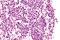
| Micrograph of a typical pulmonary carcinoid tumour. | |
| Specialty | Oncology |
Signs and symptoms
Lung carcinoids typically present with a cough or hemoptysis.[2] Findings may closely mimic malignant tumours of the lung, i.e. lung cancer.[citation needed]
Diagnosis
.jpg)
The definitive diagnosis is rendered by a microscopic examination, after excision. Typical carcinoids have cells with stippled chromatin and a moderate quantity of cytoplasm. They typically have few mitoses and lack necrosis. By definition, they are greater than 4 mm in largest dimension; smaller lesions are referred to as pulmonary carcinoid tumourlets.[citation needed]
The differential diagnosis of typical pulmonary carcinoid tumour includes: atypical pulmonary carcinoid tumour, pulmonary carcinoid tumourlet and lung adenocarcinoma.[citation needed]
 Very high magnification
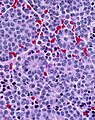 With prominent rosettes


Treatment
Typical carcinoids are usually treated with surgical excision.[citation needed]
See also
References
External links
Wikiwand in your browser!
Seamless Wikipedia browsing. On steroids.
Every time you click a link to Wikipedia, Wiktionary or Wikiquote in your browser's search results, it will show the modern Wikiwand interface.
Wikiwand extension is a five stars, simple, with minimum permission required to keep your browsing private, safe and transparent.