Top Qs
Timeline
Chat
Perspective
Anterior external arcuate fibers
From Wikipedia, the free encyclopedia

Remove ads
The anterior external arcuate fibers (ventral external arcuate fibers) vary as to their prominence: in some cases they form an almost continuous layer covering the medullary pyramids and olivary body, while in other cases they are barely visible on the surface.
This article may be confusing or unclear to readers. (April 2009) |

They arise from the cells of the gracile and cuneate nuclei, and pass forward through the reticular formation to decussate (cross over to the other side) in the middle line.
Most of them reach the surface by way of the anterior median fissure, and arch backward over the pyramid, the olive, and the lateral district of the medulla oblongata to enter the cerebellum through the inferior peduncle. The fibers are reinforced in their course by fibers emerging between the pyramid and olive.
As the fibers arch across the pyramid, they enclose a small nucleus which lies in front of and medial to the pyramid.
This is named the arcuate nucleus, and is serially continuous above with the pontine nuclei in the pons; it contains small fusiform (spindle-shaped) cells, around which some of the arcuate fibers end, and from which others arise.
Remove ads
Additional images
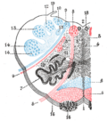 The reticular formation shown by a transverse section passing through the middle of the olive.

References
![]() This article incorporates text in the public domain from page 782 of the 20th edition of Gray's Anatomy (1918)
This article incorporates text in the public domain from page 782 of the 20th edition of Gray's Anatomy (1918)
External links
This neuroanatomy article is a stub. You can help Wikipedia by expanding it. |
Wikiwand - on
Seamless Wikipedia browsing. On steroids.
Remove ads