Top Qs
Timeline
Chat
Perspective
Deltoid ligament
Anatomical detail in the ankle From Wikipedia, the free encyclopedia

Remove ads
The deltoid ligament (or medial ligament of talocrural joint) is a strong, flat, triangular band, attached, above, to the apex and anterior and posterior borders of the medial malleolus. The deltoid ligament supports the ankle joint and also resists excessive eversion of the foot.[1] The deltoid ligament is composed of 4 fibers:
- Anterior tibiotalar ligament
- Tibiocalcaneal ligament
- Posterior tibiotalar ligament
- Tibionavicular ligament.
It consists of two sets of fibers, superficial and deep.
Remove ads
Superficial fibres
Of the superficial fibres,
- tibionavicular pass forward to be inserted into the tuberosity of the navicular bone, and immediately behind this they blend with the medial margin of the plantar calcaneonavicular ligament;
- tibiocalcaneal descend almost perpendicularly to be inserted into the whole length of the sustentaculum tali of the calcaneus;
- posterior tibiotalar from the posterior colliculus of the medial malleolus to the posteromedial surface of the talus
Remove ads
Deep fibres
The deep fibres (anterior tibiotalar) are attached from the anterior colliculus of the medial malleolus to the medial talus and medial tubercle
Coverings
The deltoid ligament is covered by the tendons of the tibialis posterior and flexor digitorum longus which are supplied by the tibial nerve (L4, L5, S1, S2, and S3).
Additional images
 Ankle joint.Deep section.
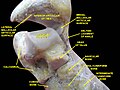 Ankle joint. Deep dissection.
 Ankle joint. Deep dissection.
 Ankle joint. Deep dissection.


References
External links
Wikiwand - on
Seamless Wikipedia browsing. On steroids.
Remove ads