Top Qs
Timeline
Chat
Perspective
Interchromatin granule
From Wikipedia, the free encyclopedia
Remove ads
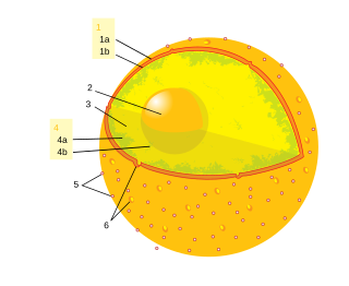
An interchromatin granule is a cluster in the nucleus of a mammal cell which is enriched in pre-mRNA splicing factors. Interchromatin granules are located in the interchromatin regions of mammal cell nuclei.[1][2][a] They usually appear as irregularly shaped structures that vary in size and number. They can be observed by immunofluorescence microscopy.[2][7]
Interchromatin granules are structures undergoing constant change, and their components exchange continuously with the nucleoplasm, active transcription sites and other nuclear locations.[2][7][8]
Research on dynamics of interchromatin granules has provided new insight into the functional organisation of the nucleus and gene expression.
Interchromatin granule clusters vary in size anywhere between one and several micrometers in diameter. They are composed of 20–25 nm granules[9] that are connected in a beaded chain fashion appearance by thin fibrils.
Interchromatin granule clusters (IGCs) may represent small nuclear ribonucleoproteins (snRNPs) that have completed their maturation process and can be supplied to nearby areas containing perichromatin fibers where splicing is taking place.[7] Other proteins, such as RNA polymerase II and certain transcription factors, as well as poly-adenylated RNA may also be present.[2] The maturation of snRNPs takes place in part in Cajal bodies, and IGCs may donate splicing factors to the snRNP.[8]
Remove ads
See also
- Cell nucleus § Splicing speckles are subnuclear structures that are enriched in pre-messenger RNA splicing factors
Notes
References
Wikiwand - on
Seamless Wikipedia browsing. On steroids.
Remove ads


