Top Qs
Timeline
Chat
Perspective
Optic vesicle
Sac that protrudes from the embryonic forebrain to form each eye From Wikipedia, the free encyclopedia

Remove ads
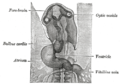
The eyes begin to develop as a pair of diverticula (pouches) from the lateral aspects of the forebrain. These diverticula make their appearance before the closure of the anterior end of the neural tube;[1][2] after the closure of the tube around the 4th week of development, they are known as the optic vesicles. Previous studies of optic vesicles suggest that the surrounding extraocular tissues – the surface ectoderm and extraocular mesenchyme – are necessary for normal eye growth and differentiation.[3]

They project toward the sides of the head, and the peripheral part of each expands to form a hollow bulb, while the proximal part remains narrow and constitutes the optic stalk, which goes on to form the optic nerve.[4][5]
Remove ads
Additional images
 Head of chick embryo of about thirty-eight hours’ incubation, viewed from the ventral surface. X 26

See also
References
External links
Wikiwand - on
Seamless Wikipedia browsing. On steroids.
Remove ads