پرونده:NMJ.jpg
From Wikipedia, the free encyclopedia
Electron_micrograph_of_neuromuscular_junction_(cross-section).jpg (۴۳۳ × ۲۸۹ پیکسل، اندازهٔ پرونده: ۹۵ کیلوبایت، نوع MIME پرونده: image/jpeg)
این پرونده در ویکیانبار موجود است. محتویات صفحهٔ توصیف آن در زیر نمایش داده میشود. |
خلاصه
| توضیحElectron micrograph of neuromuscular junction (cross-section).jpg |
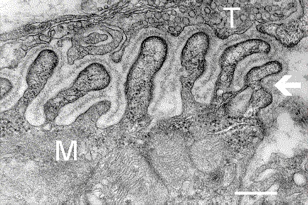
English: Electron micrograph showing a cross-section through the neuromuscular junction. T is the axon terminal, M is the muscle fiber. The arrow shows junctional folds with basal lamina. Postsynaptic densities are visible on the tips between the folds. The scale is 0.3 µm. |
| تاریخ | Originally uploaded to en.wikipedia on ۱۰ مارس ۲۰۰۶. |
| منبع | Synapse Web at the National Institute of Mental Health, National Institutes of Health; originally from en.wikipedia; description page is/was here. |
| پدیدآور | National Institute of Mental Health; originally uploaded by Nrets at en.wikipedia. |
اجازهنامه
| Public domainPublic domainfalsefalse |
This image is a work of the National Institutes of Health, part of the United States Department of Health and Human Services, taken or made as part of an employee's official duties. As a work of the U.S. federal government, the image is in the public domain.
 |
||
| این پرونده تحت قانون حق تکثیر محدودیت آزاد منتشر شده که شامل تمامی حقوق مربوطه و حقوق نزدیک به آن میشود. | ||
https://creativecommons.org/publicdomain/mark/1.0/PDMCreative Commons Public Domain Mark 1.0falsefalse
سیاهه بارگذاری اصلی
(All user names refer to en.wikipedia)
- 2006-03-10 20:07 Nrets 433×289×8 (97758 bytes) Electron micrograph showing a cross section through the neuromuscular junction. T is the axon terminal, M is the muscle fiber. The arrow shows junctional folds with basal lamina. Postsynaptic densities are visible on the tips between the folds. Scale is 0
عنوان
آیتمهایی که در این پرونده نمایش داده شدهاند
توصیفها
۱۰ مارس 2006
تاریخچهٔ پرونده
روی تاریخ/زمانها کلیک کنید تا نسخهٔ مربوط به آن هنگام را ببینید.
| تاریخ/زمان | بندانگشتی | ابعاد | کاربر | توضیح | |
|---|---|---|---|---|---|
| کنونی | ۲۲ مارس ۲۰۰۷، ساعت ۰۳:۴۱ | | ۴۳۳ در ۲۸۹ (۹۵ کیلوبایت) | Fran Rogers | {{Information |Description=Electron micrograph showing a cross section through the neuromuscular junction. T is the axon terminal, M is the muscle fiber. The arrow shows junctional folds with basal lamina. Postsynaptic densities are visible on the tips be |
کاربرد پرونده
صفحهٔ زیر از این تصویر استفاده میکند:
کاربرد سراسری پرونده
ویکیهای دیگر زیر از این پرونده استفاده میکنند:
- کاربرد در ar.wikipedia.org
- کاربرد در cs.wikipedia.org
- کاربرد در de.wikipedia.org
- کاربرد در es.wikipedia.org
- کاربرد در gl.wikipedia.org
- کاربرد در he.wikipedia.org
- کاربرد در ko.wikipedia.org
- کاربرد در pt.wikipedia.org
- کاربرد در ru.wikipedia.org
- کاربرد در uk.wikipedia.org
- کاربرد در zh.wikipedia.org
فراداده
این پرونده حاوی اطلاعات اضافهایاست که احتمالاً دوربین دیجیتال یا پویشگری که در ایجاد یا دیجیتالیکردن آن به کار رفته آن را افزوده است. اگر پرونده از وضعیت ابتداییاش تغییر داده شده باشد آنگاه ممکن است شرح و تفصیلات موجود اطلاعات تصویر را تماماً بازتاب ندهد.
| جهت | عادی |
|---|
.jpg)