ファイル:Ebola_virus_em.png
ウィキペディア フリーな encyclopedia
元のファイル (2,043 × 2,887 ピクセル、ファイルサイズ: 2.55メガバイト、MIME タイプ: image/png)
ウィキメディア・コモンズのファイルページにある説明を、以下に表示します。
|
| 解説Ebola virus em.png |
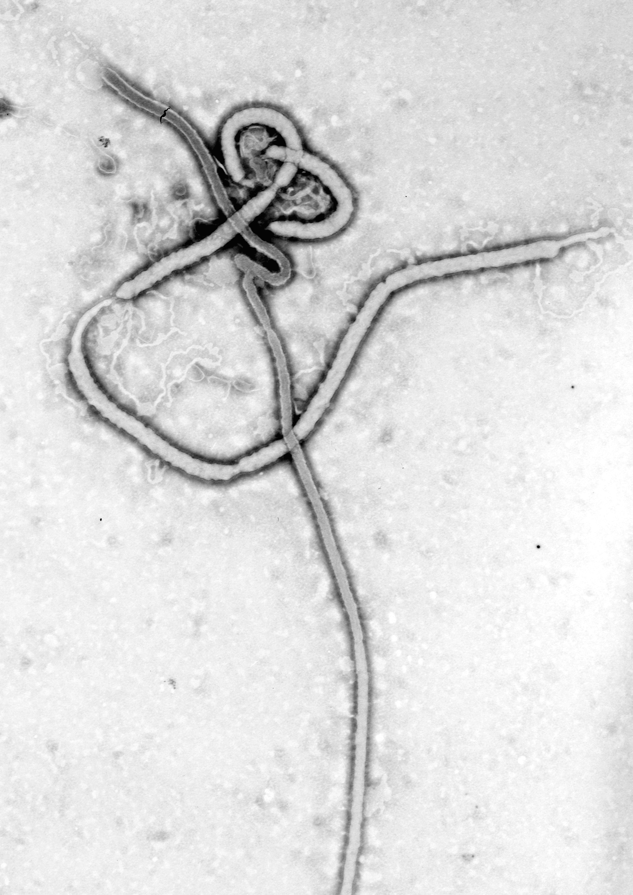
English: An electron micrograph of an Ebola viral particle showing the characteristic filamentous structure of a Filoviridae. The viral filaments can appear in images in various shapes including a 'u', '6', a coil, or branched resulting in pleomorphic particles. The filaments are reported to be between 60-80 nm in diameter, the length of a filament associated with an individual viral partial is extremely variable with Ebola particles of up to 14,000 nm in length being reported. An average length, which may represent the most infectious particles is in the region of 1000 nm.
The first electronmicrograph of Ebola was 13 October 1976 by Dr. F.A. Murphy, now at UC Davis, who was then working at the CDC. The nucleocapsid structure consists of a central channel, 20-30nm in diameter, surrounded by helically wound capsid with a diameter of 40-50nm and an interval of 5nm. 7nm glycoprotein spikes spaced 10 nm apart from each other are present within the outer envelope of the virus which is derived from the host cell membrane. Each viral particle contains one molecule of single-stranded, negative-sense RNA, which encodes the seven viral proteins.Polski: Grafika przedstawiająca wirus Ebola pochodząca z mikroskopu elektronowego ukazująca
charakterystyczną filamentową budowę tego Filowirusa. Te wirusowe filamenty występują na obrzach w różnych postaciach między innymi: "u" '6' albo sprężyny. Filamenty występują w rozmiarze 60-80 nm średnicy. Długość filamentu jest bardzo zmienna i zależna od konkretnej "cząsteczki" wirusa, w przypadku Ebola pojawiają się mające długość nawet 14,000 nm. Średnia długość, która może reprezentować wersję najbardziej zaraźliwą ma długość około 1000nm. Pierwsze zdjęcie wirusa Ebola zostało wykonane 13 Października 1976 roku przez Dr. F.A Murphy, teraz pracującego na uniwersytecie w Davis, wtedy w CDC.中文:
Source: CDC
|
||||
| 日付 | |||||
| 原典 |
|
||||
| 作者 | CDC/ Dr. Frederick A. Murphy | ||||
| 許可 (ファイルの再利用) |
|
キャプション
1976
ファイルの履歴
過去の版のファイルを表示するには、その版の日時をクリックしてください。
| 日付と時刻 | サムネイル | 寸法 | 利用者 | コメント | |
|---|---|---|---|---|---|
| 現在の版 | 2014年7月28日 (月) 09:33 | | 2,043 × 2,887 (2.55メガバイト) | Splintercellguy | Upload higher-resolution version |
| 2005年5月24日 (火) 11:22 |  | 150 × 227 (19キロバイト) | Knutux | An electron micrograph of an Ebola viral particle showing the characteristic filamentous structure of a Filoviridae. The viral filaments can appear in images in various shapes including a 'u', '6', a coil, or branched resul |
ファイルの使用状況
以下のページがこのファイルを使用しています:
グローバルなファイル使用状況
以下に挙げる他のウィキがこの画像を使っています:
- af.wikipedia.org での使用状況
- als.wikipedia.org での使用状況
- ar.wikipedia.org での使用状況
- arz.wikipedia.org での使用状況
- ast.wikipedia.org での使用状況
- bn.wikipedia.org での使用状況
- ca.wikipedia.org での使用状況
- ca.wikinews.org での使用状況
- csb.wikipedia.org での使用状況
- da.wikipedia.org での使用状況
- de.wikipedia.org での使用状況
- el.wikipedia.org での使用状況
- en.wikipedia.org での使用状況
- Wikipedia:Main Page/French
- Wikipedia:WikiProject Viruses/Templates
- Virus
- Wikipedia:Top 25 Report/July 27 to August 2, 2014
- Wikipedia:Top 25 Report/August 3 to 9, 2014
- RVSV-ZEBOV vaccine
- CAd3-ZEBOV
- 2016 in science
- Wikipedia:Top 25 Report/Records
- User:M1Abramstanbks/sandbox
- Wikipedia:In the news/Posted/May 2004
- en.wikinews.org での使用状況
- eo.wikipedia.org での使用状況
- eo.wikinews.org での使用状況
- es.wikipedia.org での使用状況
- et.wikipedia.org での使用状況
- eu.wikipedia.org での使用状況
- fi.wikiversity.org での使用状況
- fr.wikipedia.org での使用状況
- fr.wikibooks.org での使用状況
- ga.wikipedia.org での使用状況
- gl.wikipedia.org での使用状況
このファイルのグローバル使用状況を表示する。
メタデータ
このファイルには、追加情報があります (おそらく、作成やデジタル化する際に使用したデジタルカメラやスキャナーが追加したものです)。
このファイルが元の状態から変更されている場合、修正されたファイルを完全に反映していない項目がある場合があります。
| 水平方向の解像度 | 472.44dpc |
|---|---|
| 垂直方向の解像度 | 472.44dpc |
| ファイル変更日時 | 2014年7月28日 (月) 09:32 |