File:Leaf_epidermis_w_scale.jpg
From Wikipedia, the free encyclopedia
Size of this preview: 598 × 599 pixels. Other resolutions: 240 × 240 pixels | 479 × 480 pixels | 767 × 768 pixels | 1,022 × 1,024 pixels | 2,048 × 2,052 pixels.
Original file (2,048 × 2,052 pixels, file size: 1.1 MB, MIME type: image/jpeg)
| This is a file from the Wikimedia Commons. Information from its description page there is shown below. Commons is a freely licensed media file repository. You can help. |
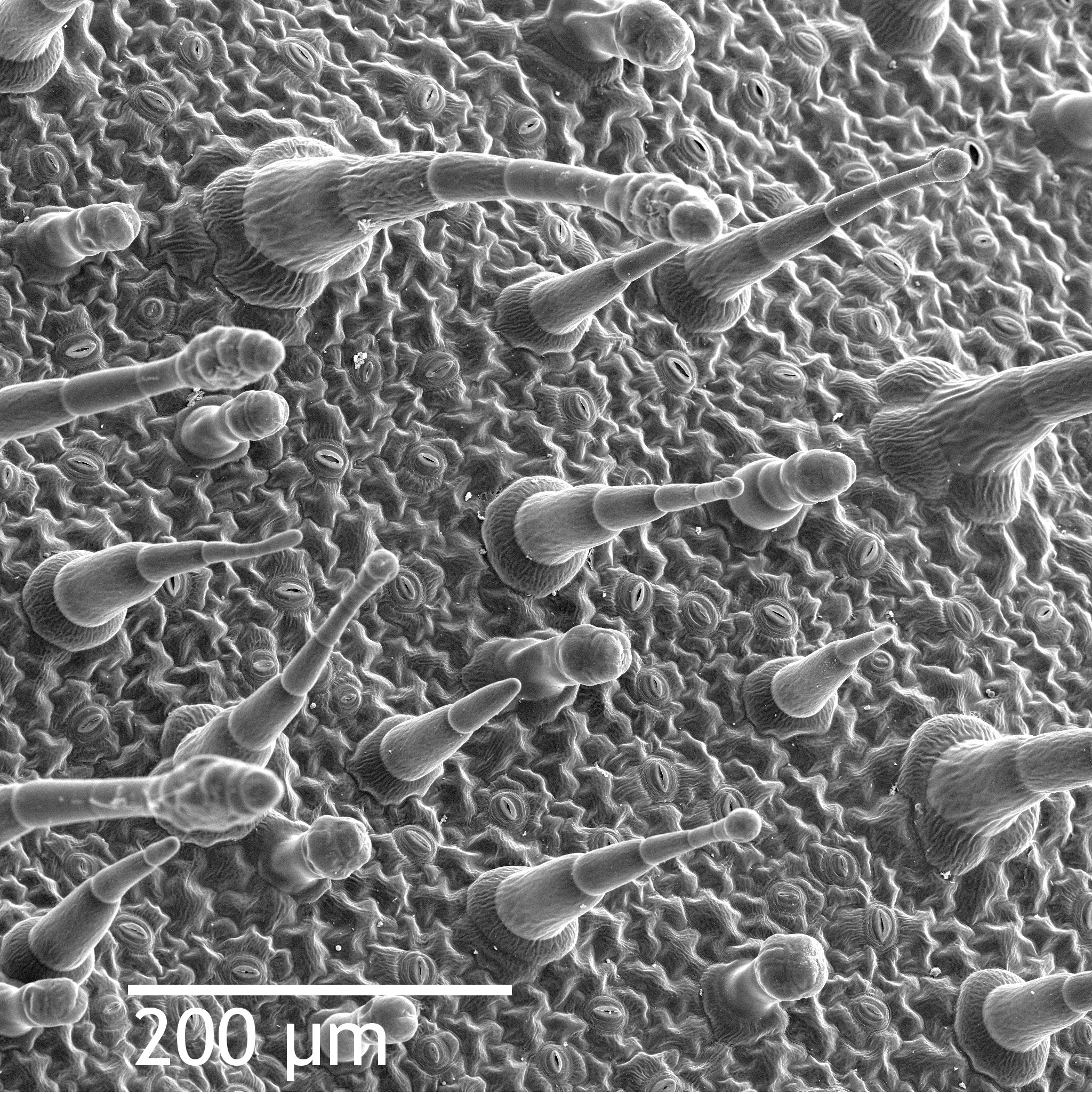
| DescriptionLeaf epidermis w scale.jpg | Scanning electron microscope image of Nicotiana alata upper leaf surface, showing tricomes and a few stomates. Instrument: ZEISS962 SEM. |
| Date | (UTC) |
| Source | |
| Author |
|
| This is a retouched picture, which means that it has been digitally altered from its original version. Modifications: Added scale, more contrast. The original can be viewed here: Leaf epidermis.jpg:
|
| Public domainPublic domainfalsefalse |
| This work has been released into the public domain by its author, Louisa Howard. This applies worldwide. In some countries this may not be legally possible; if so: Louisa Howard grants anyone the right to use this work for any purpose, without any conditions, unless such conditions are required by law. Public domainPublic domainfalsefalse |
Original upload log
This image is a derivative work of the following images:
- File:Leaf_epidermis.jpg licensed with PD-author
- 2008-06-21T18:26:19Z Mangostar 2048x2073 (3038992 Bytes) {{Information |Description=Scanning electron microscope image of Nicotiana alata upper leaf surface, showing tricomes and a few stomates. Instrument: ZEISS962 SEM. |Source=http://remf.dartmouth.edu/images/NicotianaLeafSEM/nic
Uploaded with derivativeFX
Captions
Add a one-line explanation of what this file represents
Items portrayed in this file
depicts
11 March 2010
File history
Click on a date/time to view the file as it appeared at that time.
| Date/Time | Thumbnail | Dimensions | User | Comment | |
|---|---|---|---|---|---|
| current | 01:59, 11 March 2010 | | 2,048 × 2,052 (1.1 MB) | Laitr Keiows | {{Information |Description=Scanning electron microscope image of Nicotiana alata upper leaf surface, showing tricomes and a few stomates. Instrument: ZEISS962 SEM. |Source=*File:Leaf_epidermis.jpg |Date=2010-03-11 01:58 (UTC) |Author=*[[:File:Leaf_e |
File usage
The following page uses this file:
Global file usage
The following other wikis use this file:
- Usage on ar.wikipedia.org
- Usage on bn.wikipedia.org
- Usage on bs.wikipedia.org
- Usage on en.wikipedia.org
- Usage on en.wikiversity.org
- Usage on fr.wikipedia.org
- Usage on gl.wikipedia.org
- Usage on gv.wikipedia.org
- Usage on ht.wikipedia.org
- Usage on id.wikipedia.org
- Usage on it.wikipedia.org
- Usage on ja.wikipedia.org
- Usage on kk.wikipedia.org
- Usage on la.wikipedia.org
- Usage on lv.wikipedia.org
- Usage on ml.wikipedia.org
- Usage on ms.wikipedia.org
- Usage on ru.wikipedia.org
- Usage on sl.wikipedia.org
- Usage on vi.wikipedia.org
- Usage on zh.wikipedia.org
Metadata
This file contains additional information, probably added from the digital camera or scanner used to create or digitize it.
If the file has been modified from its original state, some details may not fully reflect the modified file.
| _error | 0 |
|---|
Retrieved from "https://simple.wikipedia.org/wiki/File:Leaf_epidermis_w_scale.jpg"