Fitxer:Fundus_of_patient_with_retinitis_pigmentosa,_mid_stage.jpg
From Wikipedia, the free encyclopedia
Mida d'aquesta previsualització: 699 × 599 píxels. Altres resolucions: 280 × 240 píxels | 560 × 480 píxels | 871 × 747 píxels.
Fitxer original (871 × 747 píxels, mida del fitxer: 107 Ko, tipus MIME: image/jpeg)
| Aquest fitxer i la informació mostrada a continuació provenen del dipòsit multimèdia lliure Wikimedia Commons. |
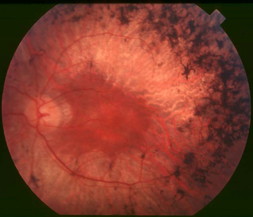
| DescripcióFundus of patient with retinitis pigmentosa, mid stage.jpg |
English: Figure 2. Fundus of patient with retinitis pigmentosa, mid stage (Bone spicule-shaped pigment deposits are present in the mid periphery along with retinal atrophy, while the macula is preserved although with a peripheral ring of depigmentation. Retinal vessels are attenuated.) Hamel Orphanet Journal of Rare Diseases 2006 1:40 doi:10.1186/1750-1172-1-40 |
| Data | |
| Font | Retinitis pigmentosa by Christian Hamel |
| Autor | Christian Hamel |
| Permís (Com reutilitzar aquest fitxer) |
© 2006 Hamel; licensee BioMed Central Ltd. This is an Open Access article distributed under the terms of the Creative Commons Attribution License (https://creativecommons.org/licenses/by/2.0), which permits unrestricted use, distribution, and reproduction in any medium, provided the original work is properly cited. |
Aquest fitxer està disponible sota la llicència Creative Commons Reconeixement 2.0 Genèrica.
- Sou lliure de:
- compartir – copiar, distribuir i comunicar públicament l'obra
- adaptar – fer-ne obres derivades
- Amb les condicions següents:
- reconeixement – Heu de donar la informació adequada sobre l'autor, proporcionar un enllaç a la llicència i indicar si s'han realitzat canvis. Podeu fer-ho amb qualsevol mitjà raonable, però de cap manera no suggereixi que l'autor us dóna suport o aprova l'ús que en feu.
Llegendes
Afegeix una explicació d'una línia del que representa aquest fitxer
Fundus of patient with retinitis pigmentosa
Elements representats en aquest fitxer
representa l'entitat
Historial del fitxer
Cliqueu una data/hora per veure el fitxer tal com era aleshores.
| Data/hora | Miniatura | Dimensions | Usuari/a | Comentari | |
|---|---|---|---|---|---|
| actual | 12:17, 2 des 2017 | | 871 × 747 (107 Ko) | Doc James | Cropped 27 % horizontally and 7 % vertically using CropTool with precise mode. |
| 15:52, 22 set 2009 |  | 1.200 × 799 (126 Ko) | CopperKettle | {{Information |Description={{en|1=Figure 2. Fundus of patient with retinitis pigmentosa, mid stage (Bone spicule-shaped pigment deposits are present in the mid periphery along with retinal atrophy, while the macula is preserved although with a peripheral |
Ús del fitxer
La pàgina següent utilitza aquest fitxer:
Ús global del fitxer
Utilització d'aquest fitxer en altres wikis:
- Utilització a ar.wikipedia.org
- Utilització a bs.wikipedia.org
- Utilització a da.wikipedia.org
- Utilització a en.wikipedia.org
- Utilització a en.wikiversity.org
- Utilització a es.wikipedia.org
- Utilització a eu.wikipedia.org
- Utilització a fa.wikipedia.org
- Utilització a fi.wikipedia.org
- Utilització a fr.wikipedia.org
- Utilització a he.wikipedia.org
- Utilització a hy.wikipedia.org
- Utilització a it.wikipedia.org
- Utilització a ko.wikipedia.org
- Utilització a la.wikipedia.org
- Utilització a or.wikipedia.org
- Utilització a outreach.wikimedia.org
- Utilització a pl.wikipedia.org
- Utilització a pt.wikipedia.org
- Utilització a ru.wikipedia.org
- Utilització a sl.wikipedia.org
- Utilització a sr.wikipedia.org
- Utilització a sv.wikipedia.org
- Utilització a th.wikipedia.org
- Utilització a tr.wikipedia.org
- Utilització a tt.wikipedia.org
- Utilització a uk.wikipedia.org
- Utilització a vi.wikipedia.org
Vegeu més usos globals d'aquest fitxer.