File:Mitochondria,_mammalian_lung_-_TEM.jpg
From Wikipedia, the free encyclopedia
No higher resolution available.
Mitochondria,_mammalian_lung_-_TEM.jpg (640 × 480 pixels, file size: 96 KB, MIME type: image/jpeg)
| This is a file from the Wikimedia Commons. Information from its description page there is shown below. Commons is a freely licensed media file repository. You can help. |
Summary
| DescriptionMitochondria, mammalian lung - TEM.jpg |
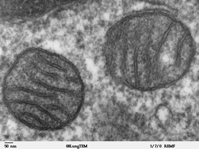
Transmission electron microscope image of a thin section cut through an area of mammalian lung tissue. The high magnification image shows two mitochondria. JEOL 100CX TEM |
| Source | |
| Author | Louisa Howard |
| Permission (Reusing this file) |
PD |
Licensing
| Public domainPublic domainfalsefalse |
| This work has been released into the public domain by its author, Louisa Howard. This applies worldwide. In some countries this may not be legally possible; if so: Louisa Howard grants anyone the right to use this work for any purpose, without any conditions, unless such conditions are required by law. Public domainPublic domainfalsefalse |
Captions
Add a one-line explanation of what this file represents
Mammalian Lung tissue
Митохондрия под микроскопом
h
Items portrayed in this file
depicts
image/jpeg
File history
Click on a date/time to view the file as it appeared at that time.
| Date/Time | Thumbnail | Dimensions | User | Comment | |
|---|---|---|---|---|---|
| current | 15:09, 16 May 2008 | | 640 × 480 (96 KB) | Vojtěch Dostál | Reverted to version as of 15:37, 5 October 2006, my fault |
| 12:51, 16 May 2008 |  | 640 × 433 (84 KB) | Vojtěch Dostál | {{Information |Description=Transmission electron microscope image of a thin section cut through an area of mammalian lung tissue. The high magnification image shows a mitochondria. JEOL 100CX TEM |Source= * http://remf.dartmouth.edu/imagesindex.html * h | |
| 12:47, 16 May 2008 |  | 640 × 453 (86 KB) | Vojtěch Dostál | {{Information |Description=Transmission electron microscope image of a thin section cut through an area of mammalian lung tissue. The high magnification image shows a mitochondria. JEOL 100CX TEM |Source= * http://remf.dartmouth.edu/imagesindex.html * h | |
| 15:37, 5 October 2006 |  | 640 × 480 (96 KB) | Patho | {{Information |Description=Transmission electron microscope image of a thin section cut through an area of mammalian lung tissue. The high magnification image shows a mitochondria. JEOL 100CX TEM |Source= * http://remf.dartmouth.edu/imagesindex.html * h |
File usage
The following pages on the English Wikipedia use this file (pages on other projects are not listed):
Global file usage
The following other wikis use this file:
- Usage on ar.wikipedia.org
- Usage on az.wiktionary.org
- Usage on be.wikipedia.org
- Usage on bg.wikipedia.org
- Usage on bn.wikipedia.org
- Usage on br.wikipedia.org
- Usage on bs.wikipedia.org
- Usage on ca.wikipedia.org
- Usage on cdo.wikipedia.org
- Usage on da.wikipedia.org
- Usage on de.wikibooks.org
- Usage on el.wikipedia.org
- Usage on en.wikibooks.org
- Usage on en.wikiversity.org
- User:Jtwsaddress42/Projects/Project 1
- User:Jtwsaddress42/Projects/Project 1/Parts
- User:Jtwsaddress42/Projects/Project 1/Parts/Part 3
- User:Jtwsaddress42/Projects/Project 1/Chapters/Chapter 10
- User:Jtwsaddress42/Projects/Project 1/Sections/Chapter 10/Phase II - The Oxygen Crisis and the Rise of the Aerobic Bioshphere (1.9-0.95 bya)
- User:Jtwsaddress42/Clade
- User:Jtwsaddress42/Clade/Gracilicutes to Proteobacteria
- Usage on en.wiktionary.org
- Usage on es.wikipedia.org
- Usage on et.wikipedia.org
- Usage on eu.wikipedia.org
- Usage on ext.wikipedia.org
- Usage on fa.wikipedia.org
- Usage on fr.wikipedia.org
- Usage on ga.wikipedia.org
- Usage on gl.wikipedia.org
- Usage on gv.wikipedia.org
- Usage on he.wikipedia.org
View more global usage of this file.
Metadata
This file contains additional information, probably added from the digital camera or scanner used to create or digitize it.
If the file has been modified from its original state, some details may not fully reflect the modified file.
| _error | 0 |
|---|