Top Qs
Timeline
Chat
Perspective
Aneurysmal bone cyst
Benign bone tumor consisting of blood From Wikipedia, the free encyclopedia

Remove ads
Aneurysmal bone cyst (ABC) is a non-cancerous bone tumor composed of multiple varying sizes of spaces in a bone which are filled with blood.[1][4] The term is a misnomer, as the lesion is neither an aneurysm nor a cyst.[5] It generally presents with pain and swelling in the affected bone.[1] Pressure on neighbouring tissues may cause compression effects such as neurological symptoms.[1]
The cause is unknown.[1] Diagnosis involves medical imaging.[1] CT scan and X-ray show lytic expansion lesions with clear borders.[1] MRI reveals fluid levels.[1]
Treatment is usually by curettage, bone grafting or surgically removing the part of bone.[2] 20–30% may recur, usually in the first couple of years after treatment, particularly in children.[2]
It is rare.[3] The incidence is around 0.15 cases per one million per year.[1] Aneurysmal bone cyst was first described by Jaffe and Lichtenstein in 1942.[5][6]
Remove ads
Signs and symptoms
The afflicted may have relatively small amounts of pain that will quickly increase in severity over a time period of 6–12 weeks. The skin temperature around the bone may increase, a bony swelling may be evident, and movement may be restricted in adjacent joints.[7]
Spinal lesions may cause quadriplegia and patients with skull lesions may have headaches.[citation needed]
Sites
Commonly affected sites are metaphyses of vertebra, flat bones, femur and tibia.[8] Approximate percentages by sites are as shown:[citation needed]
- Skull and mandible (4%)
- Spine (16%)
- Clavicle and ribs (5%)
- Upper extremity (21%)
- Pelvis and sacrum (12%)
- Femur (13%)
- Lower leg (24%)
- Foot (3%)
Remove ads
Causes
Aneurysmal bone cyst has been widely regarded a reactive process of uncertain cause since its initial description by Jaffe and Lichtenstein in 1942. Many hypotheses have been proposed to explain the cause and pathogenesis of aneurysmal bone cyst, and until very recently the most commonly accepted idea was that aneurysmal bone cyst was the consequence of an increased venous pressure and resultant dilation and rupture of the local vascular network. However, studies by Panoutsakopoulus et al. and Oliveira et al. uncovered the clonal neoplastic nature of aneurysmal bone cyst. Primary cause has been regarded arteriovenous fistula within bone.[9]
The lesion may arise de novo or may arise secondarily within a pre-existing bone tumor, because the abnormal bone causes changes in hemodynamics. An aneurysmal bone cyst can arise from a pre-existing chondroblastoma, a chondromyxoid fibroma, an osteoblastoma, a giant cell tumor, or fibrous dysplasia. A giant cell tumor is the most common cause, occurring in 19–39% of cases. Less frequently, it results from some malignant tumors, such as osteosarcoma, chondrosarcoma, and hemangioendothelioma.[citation needed]
Remove ads
Pathology

Histologically, they are classified in two variants.[citation needed]
- The classic (or standard) form (95%) has blood filled clefts among bony trabeculae. Osteoid tissue is found in stromal matrix.
- The solid form (5%) shows fibroblastic proliferation, osteoid production and degenerated calcifying fibromyxoid elements.
According to Buraczewski and Dabska, the development of the aneurysmal bone cyst follows three stages.[5]
They can also be associated with a TRE17/USP6 translocation.[10]
Aneurysmal bone cysts may be intraosseous, staying inside of the bone marrow. Or they may be extraosseous, developing on the surface of the bone, and extending into the marrow. A radiograph will reveal a soap bubble appearance.[citation needed]
Diagnosis
X-ray and CT scan show lytic expansion lesions with clear borders.[1] Expansion of cortex gives the lesion a balloon-like appearance. Larger lesions may appear septated.[11] MRI reveals fluid levels.[1] Bone scan shows outer radiotracer uptake, with a central dark area.[1][11]
Differential diagnosis
Following conditions are excluded before diagnosis can be confirmed:[12]
- Unicameral bone cyst
- Giant cell tumor
- Telangiectatic osteosarcoma
- Secondary aneurysmal bone cyst
Remove ads
Treatment
Curettage is performed on some people,[13] and is sufficient for inactive lesions. The recurrence rate with curettage is significant in active lesions, and marginal resection has been advised. Liquid nitrogen, phenol, methyl methacrylate are considered for use to kill cells at margins of resected cyst.[9]
Prognosis
20–70% recur after curettage.[1]
Epidemiology
It is rare.[3] The incidence is around 0.15 cases per one million per year.[1] 80% occur in people age less than 20 years.[1] It is second most common tumor of spine and most common benign tumor of pelvis in pediatric population.[9] Males and females are equally affected.[1]
Additional images
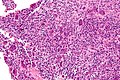 High magnification micrograph of an aneurysmal bone cyst
 Intermediate magnification micrograph of an aneurysmal bone cyst


See also
References
External links
Wikiwand - on
Seamless Wikipedia browsing. On steroids.
Remove ads