In suspected immunologic disease:[4] Fixation for immunofluorescence, with for example Michel's solution.[3]For details, see immunofluorescense of skin tissues
If suspected bacterial and fungal microorganisms, consider Gram stain and Gomori methenamine silver stain.[2]
Microscopic evaluation
Summarize
Perspective
One approach is to classify into mainly either of the following, primarily based on depth of involvement:[2]
Epidermis, papillary dermis, and superficial vascular plexus:
Vesiculobullous lesions
Pustular dermatosis
Non vesicullobullous, non-pustular
With epidermal changes
Without epidermal changes. These characteristically have a superficial perivascular inflammatory infiltrate, and can be classified by type of cell infiltrate:[2]
Lymphocytic (most common)
Lymphoeosinophilic
Lymphoplasmacytic
Mast cell
Lymphohistiocytic
Neutrophilic
Continue in corresponding section:
Non vesicullobullous, non-pustular lesions with epidermal changes
Spongiotic dermatitis
It is characterized by epithelial intercellular edema.[2]
Focal, usually mild, spongiosis with overlying scale crust, with a few neutrophils
The crust is often centered on a follicle
The papillary dermis is generally mildly edematous
Dilated blood vessels in the superficial vascular plexus
Mild superficial perivascular infiltrate of lymphocytes, histiocytes and occasional neutrophils. There is some exocytosis of inflammatory cells but not as prominent as in nummular dermatitis
Presence of scaling crusts in a folliculocentric distribution, distinguishes from psoriasis.
Close
In addition to above, an unspecific spongiotic dermatitis can be consistent with nummular dermatitis, dyshidrotic dermatitis, Id reaction, dermatophytosis, miliaria, Gianotti-Crosti syndrome and pityriasis rosea.[2][notes 2]
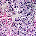
Mild inflammatory cell infiltrate along the dermoepidermal junction (black arrow in image)
Vacuolization within the basal keratinocytes (white arrow in image)
Often necrotic, predominantly basal, individual keratinocytes, manifesting as colloid or Civatte bodies
Acute graft-versus-host-disease
Vacuolar alteration of various severity, from focal or diffuse vacuolation of the basal keratinocytes (grade I), to separation at the dermoepidermal junction (grade III)
Hyperkeratosis, atrophic epidermis, sclerosis of dermis and dermal lymphocytes.[7]
Erythema multiforme
Lupus erythematosis
Typical findings in systemic lupus erythematosus:[8]
Fibrinoid necrosis at the dermoepidermal junction
Liquefactive degeneration and atrophy of the epidermis
Mucin deposition in the reticular dermis
Edema, small hemorrhages
Mild and mainly lymphocytic infiltrate in the upper dermis
Fibrinoid material in the dermis around capillary blood vessels, on collagen and in the interstitium
In non-bullous cases, perivascular and interstitial neutrophils are sometimes present in the upper dermis, with damage to blood vessels
Close
An interface dermatitis with vacuolar alteration, not otherwise specified, may be caused by viral exanthems, phototoxic dermatitis, acute radiation dermatitis, erythema dyschromicum perstans, lupus erythematosus and dermatomyositis.[2]
Interface dermatitis with lichenoid inflammation
More information Main conditions, Characteristics ...
In the papillary dermis: a confluent, band-like, dense inflammation of mainly small lymphocytes and a few histiocytes, along or hugging the dermoepidermal junction.
Often vacuolar degeneration of basal keratinocytes and apoptotic bodies (colloid or Civatte bodies).
Lichen planus
Irregular epidermal hyperplasia with a jagged "sawtooth" appearance, compact hyperkeratosis or orthokeratosis, foci of wedge-shaped hypergranulosis, basilar vacuolar degeneration, slight spongiosis in the spinous layer, and squamatization. The dermal papillae between the elongated rete ridges are frequently dome shaped. Necrotic keratinocytes can be observed in the basal layer of the epidermis and at the dermal-epidermal junction. Eosinophilic remnants of anucleate apoptotic basal cells may also be found in the dermis and are referred to as "colloid or civatte bodies". Whickham striae are usually seen in the areas of hypergranulosis. Vacuolar degeneration at the basal layer may be noted leading to focal subepidermal clefts (Max Joseph spaces). Squamatization occurs as a result of maturation and flattening of cells in the basal layer. It happens in areas of marked hypergranulosis with prominence of the sawtooth pattern of rete ridges. Wedge-shaped hypergranulosis can occur in the eccrine ducts (acrosyringia) or hair follicles (acrotrichia). In the hypertrophic subtype, the associated hyperkeratosis, parakeratosis, hypergranulosis, papillomatosis, acanthosis, and hyperplasia markedly increased with thicker collagen bundles forming in the dermis. Moreover, the rete ridges are more elongated and rounded as opposed to the typical sawtooth pattern. In atrophic LP, loss of the rete ridges and dermal fibrosis is prominent. In vesiculobullous LP, the disease progression is quicker. Hence, some of the distinctive features such as hyperkeratosis, hypergranulosis, or dense lymphocytic dermal-epidermal infiltrate may not be present. LP lesion may resolve with residual hyperpigmentation caused by a persistent increase in the number of melanophages in the papillary dermis.[9]
Lichenoid drug reaction
Can virtually be indistinguishable from cutaneous LP both clinically and histopathologically.
Typically, lesions have a photodistribution in the absence of oral mucosal involvement.[9]
Characteristically parakeratosis, a dermal eosinophilic infiltrate, and a perivascular lymphocytic infiltrate affecting the reticular dermis.
Epidermal changes are less common in lichenoid drug eruptions when compared to classic lichen planus. However, a higher concentration of necrotic keratinocyte and eosinophils in the infiltrate can be helpful in distinguishing lichenoid drug reaction from cutaneous lichen planus. A lengthy interval between the commencement of drug therapy and the onset of lesions does not exclude a diagnosis of lichenoid drug reaction. Resolution of the lesions often occurs within weeks to months after discontinuation of the offending drug.[9]
Lichen nitidus
Localized granulomatous lymphohistiocytic infiltrate in an expanded dermal papilla
Thinning of overlying epidermis and downward extension of the rete ridges at the lateral margin of the infiltrate, resulting in a typical "claw clutching a ball" appearance.[10]
Lichen amyloidosis
Presence of amyloid, possibly with direct immunofluorescence and Congo red staining.[11]
Congo red.
Close
Interface dermatitis with lichenoid inflammation, not otherwise specified, can be caused by lichen planus-like keratosis, lichenoid actinic keratosis, lichenoid lupus erythematosus, lichenoid GVHD (chronic GVHD), pigmented purpuric dermatosis, pityriasis rosea, and pityriasis lichenoides chronica.[2] Unusual conditions that can be associated with a lichenoid inflammatory cell infiltrate are HIV dermatitis, syphilis, mycosis fungoides, urticaria pigmentosa, and post-inflammatory hyperpigmentation.[2] In cases of post-inflammatory hyperpigmentation, it is important to exclude potentially harmful mimics such as a regressed melanocytic lesion or lichenoid pigmented actinic keratosis.[2]
Psoriaform dermatitis
Examining multiple deeper levels is recommended if initial cuts do not correlate well with the clinical history.[2]
Often: Thinning of epidermal cells overlying the tips of dermal papillae (suprapapillary plates), and dilated, tortuous blood vessels within these papillae
Further histopathologic diagnosis is performed by the following parameters:
Perivascular location. Mast cells are relatively sparse, potentially demonstrated with special stains, preferably tryptase stain. Extravasated erythrocytes are present in about 50% of the cases. No vasculitis.[14]
Dermal edema [solid arrows in (A,B)] and a sparse superficial predominantly perivascular and interstitial infiltrate of lymphocytes and eosinophils without signs of vasculitis (dashed arrow).[15]
A lesion with superficial lymphocytic infiltrate without additional histopathologic characteristics can be due to for example drug reactions and insect bites.[2][notes 2]
Close
Lymphoeosinophilic infiltrate
More information Main conditions, Characteristics ...
Perivascular location. Mast cells are relatively sparse, potentially demonstrated with special stains, preferably tryptase stain. Extravasated erythrocytes are present in about 50% of the cases. No vasculitis.[14]
Dermal edema (solid arrows) and a sparse superficial predominantly perivascular and interstitial infiltrate of lymphocytes and eosinophils (dashed arrow)
Prevesicular stage of bullous pemphigoid
Image at right shows influx of inflammatory cells including eosinophils and neutrophils in the dermis (solid arrow) and blister cavity (dashed arrows), and deposition of fibrin (asterisks).[15] However, the diagnosis of bullous pemphigoid consist of at least 2 positive results out of 3 criteria:[19]
Pruritus and/or predominant cutaneous blisters
Linear IgG and/or C3c deposits (in an n- serrated pattern) by direct immunofluorescence microscopy (DIF)
Positive epidermal side staining by indirect immunofluorescence microscopy on human salt-split skin (IIF SSS) on a serum sample.
A lesion with superficial lymphoeosinophilic infiltrate without additional histopathologic characteristics can be due to for example drug reactions and insect bites.[2][notes 2]
Close
Lymphoplasmacytic infiltrate
More information Main conditions, Characteristics ...
Typically enlarged, dilated capillaries and venules located in the upper dermis, angulated telangiectasias, perivascular and perifollicular lymphocytic infiltration, and superficial dermal edema.[20]
Psoriasiform hyperplasia with superficial neutrophils
Lichenoid tissue reaction, epidermal apoptosis and exocytosis of neutrophils
Superficial and deep chronic infiltrate in the dermis
Often numerous plasma cells in about 1/3 of cases
Often endothelial swelling.
Erythema migrans
Typically a superficial and deep perivascular lymphocytic infiltrate.[22] Plasma cells are typically located at the periphery of the lesion, whereas eosinophils are in the center.[22]
The patch stage typically shows irregular proliferation of jagged vascular channels in the dermis below an integral epidermis. The so-called promontory sign is sometimes found in patch stage lesions and denotes vascular spaces surrounding pre-existing blood (see image).[23]
A lesion with superficial lymphoplasmacytic infiltrate without additional histopathologic characteristics can be due to for example trauma, ulceration, scar and early cutaneous connective tissue diseases.[2][notes 2]
Close
Mastocytosis
More information Main conditions, Characteristics ...
Includes the rare disease of primary mastocytosis.[2][notes 2]
Close
Lymphohistiocytic infiltrate
Leprosy
These include bacterial infections including leprosy, and the sample should therefore be stained with Ziel-Neelsen, acid fast stains, Gomori methenamine silver, PAS, and Fite stains.[2] If negative, an unspecific lymphohistocytic dermatosis may be caused by drug reactions and viral infections.[2][notes 2]
Neutrophilic infiltrate
More information Main conditions, Characteristics ...
Subepidermal vesicles and blisters associated with accumulation of neutrophils at the papillary tips.[24]
Sometimes presence of eosinophils, giving an appearance similar to bullous pemphigoid.[24]
The histopathology is unspecific in approximately 35%–40% of the cases,[24] and direct immunofluorescence is needed, showing deposition of IgA in the papillary dermis in a granular or fibrillar pattern.[25]
Early febrile neutrophilic dermatosis (Sweet's syndrome)
Neutrophilic and lymphohistiocytic infiltrate and edema.[27]
Connective tissue disorders
Usually associated with epidermal changes.[2] (Systemic lupus erythematosis pictured)
Cutaneous small-vessel vasculitis
Neutrophils with nuclear dust (dashed arrows in image), with high affinity for postcapillary venules.[28]
Features of vascular injury: fibrinoid necrosis (asterisks) and erytrocyt extravasation (solid arrows)
Close
Multinucleated giant cells
Suture granuloma, with multinucleated giant cells surrounding (grey) suture material.
Foreign bodies indicate a foreign body granuloma.
Specific forms of multinucleated giant cells include the Touton giant cell, which contains a ring of nuclei surrounding a central homogeneous cytoplasm, with foamy cytoplasm surrounding the nuclei.[29][30] The central cytoplasm (surrounded by the nuclei) may be both amphophilic and eosinophilic.[31]
In "not otherwise specified" cases, a diagnosis may be reached by a review of the medical history and physical examination, based upon the potential conditions at hand.


.jpg)


_on_the_soles_of_the_feet._Plantar_lesions-CDC.jpg)


 Touton giant cell
.jpg)