Top Qs
Timeline
Chat
Perspective
Interpeduncular fossa
Brain segment From Wikipedia, the free encyclopedia

Remove ads
The interpeduncular fossa is a deep depression of the ventral surface of the midbrain between the two cerebal crura.[1][2][3] It has been found in humans and macaques, but not in rats or mice, showing that this is a relatively new evolutionary region.[4]

Remove ads
Structure
The interpeduncular fossa is a somewhat rhomboid-shaped area of the base of the brain.[5]
Features
The lateral wall of the interpeduncular fossa bears a groove - the oculomotor sulcus - from which[6] rootlets of the oculomotor nerve emerge from the substance of the brainstem and aggregate into a single fascicle.[3][6]
Anatomical relations
The ventral tegmental area lies at the depth of the interpeduncular fossa.[3]
Boundaries
The interpeduncular fossa is in front by the optic chiasma, behind by the antero-superior surface of the pons, antero-laterally by the converging optic tracts, and postero-laterally by the diverging cerebral peduncles.[5]
The floor of interpeduncular fossa, from behind forward,[citation needed] are the posterior perforated substance,[2] corpora mamillaria, tuber cinereum, infundibulum, and pituitary gland.[citation needed]
Contents
Contents of interpeduncular fossa include oculomotor nerve, and circle of Willis.[citation needed]
The basal veins pass alongside the interpeduncular fossa before joining the great cerebral vein.[7]
Remove ads
Clinical significance
The most common locations for neurocutaneous melanosis have occurred along the interpeduncular fossa, ventral brainstem, upper cervical cord, and ventral lumbosacral cord.[8]
See also
Additional images
 Human brainstem anterior view
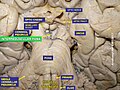 Interpeduncular fossa. Cerebrum. Deep dissection. Inferior dissection.


References
External links
Wikiwand - on
Seamless Wikipedia browsing. On steroids.
Remove ads