Top Qs
Timeline
Chat
Perspective
Lateral condyle of femur
From Wikipedia, the free encyclopedia

Remove ads
The lateral condyle is one of the two projections on the lower extremity of the femur. The other one is the medial condyle. The lateral condyle is the more prominent and is broader both in its front-to-back and transverse diameters.

Remove ads
Clinical significance
The most common injury to the lateral femoral condyle is an osteochondral fracture combined with a patellar dislocation.[1] The osteochondral fracture occurs on the weight-bearing portion of the lateral condyle. Typically, the condyle will fracture (and the patella may dislocate) as a result of severe impaction from activities such as downhill skiing and parachuting.[1]
Open reduction and internal fixation surgery is typically used to repair an osteochondral fracture. For a AO Type B1[2][circular reference] partial articular fracture of the lateral condyle, interfragmentary lag screws are used to secure the bone back together. Supplementation of buttress screws or a buttress plate is used if the fracture extends to the proximal metaphysis or distal diaphysis.[3]
Remove ads
Additional images
 Knee diagram
 Right knee-joint, from the front, showing interior ligaments.
 Muscles of the back of the leg. Deep layer.
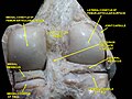 Right knee in extension. Deep dissection. Posterior view.


References
External links
Wikiwand - on
Seamless Wikipedia browsing. On steroids.
Remove ads