Top Qs
Timeline
Chat
Perspective
Myomesin-2
Protein-coding gene in the species Homo sapiens From Wikipedia, the free encyclopedia
Remove ads
Myomesin-2, also known as M-protein is a protein that in humans is encoded by the MYOM2 gene.[5] M-protein is expressed in adult cardiac muscle and fast skeletal muscle, and functions to stabilize the three-dimensional arrangement of proteins comprising M-band structures in a sarcomere.
Remove ads
Structure
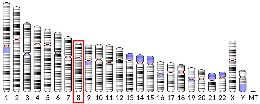
Human M-protein is 165.0 kDa and 1465 amino acids in length.[6] MYOM2 is localized to the human chromosome 8p23.3.[7] M-protein belong to the superfamily of cytoskeletal proteins having immunoglobulin/fibronectin repeats; M-protein contains two immunoglobulin C2-type repeats in the N-terminal region, five fibronectin type III repeats in the central region, and an additional four immunoglobulin C2-type repeats in the C-terminal region.[8] M-protein is expressed only in striated muscle, including fast skeletal muscle and cardiac muscle.[9][10][11]
Remove ads
Function
Summarize
Perspective
M-protein exhibits a different pattern of expression in cardiac and skeletal muscle, as well as fast- versus slow-skeletal muscle during development, suggesting different regulatory mechanisms for expression quantity and temporal appearance. In cardiac muscle, expression of M-protein continues to increase from neonatal to adult; however, in skeletal muscle, M-protein mRNA expression is biophasic.[12] M-protein is initially present in both slow- and fast-skeletal muscle embryonic fibers, then M-protein is suppressed in slow fibers.[11][13] The embryonic splice variant of myomesin, termed EH-myomesin, is expressed in a complementary pattern with M-protein during development in higher vertebrates.[14] It was also shown that the mRNA expression of M-protein is exquisitely sensitive to thyroid hormone (T3); M-protein expression, but not MYOM1 or its variant, EH-myomesin, was rapidly reduced by T3 in vivo and in vitro. The M-protein promoter is responsive to T3, and was suggested to contain thyroid hormone response elements near the transcriptional start point.[15]
The giant protein titin, together with its associated proteins, interconnects the major structure of sarcomeres, the M bands and Z discs. The C-terminal end of the titin string extends into the M line, where it binds tightly to M-band constituents MYOM1 and M-protein, of apparent molecular masses of 190 kD and 165 kD, respectively. M-protein functions to stabilize the M-line cross-linking titin and myosin; the central portion of M-protein is around the M1-line, and the N-terminal and C-terminal regions are arranged along thick filaments.[10]
An animal model of thyroid hormone (T3)-induced cardiac hypertrophy showed that T3 rapidly reduced levels of M-protein; and siRNA reduction of M-protein in neonatal cardiomyocytes showed that the absence of M-protein causes significant contractile dysfunction (77% reduction in contraction velocity), thus illuminating the importance of M-protein for normal sarcomere function.[15]
M-protein can be post-translationally modified in vivo. M-protein fragments generated via cleavage by matrix metalloproteinase 2 in left ventricular myocardium have been identified as a factor in the development of pulmonary hypertension and ascites in broiler chickens.[16] Another study demonstrated that M-protein is S-thiolated during post-ischemic reperfusion.[17] It was also determined that domains Mp2 to Mp3 in M-protein binds myosin, and this specific interaction can be regulated by phosphorylation.[18]
Remove ads
Clinical Significance
This section is empty. You can help by adding to it. (June 2015) |
Interactions
M-protein interacts with:
References
Further reading
Wikiwand - on
Seamless Wikipedia browsing. On steroids.
Remove ads


