Top Qs
Timeline
Chat
Perspective
Movat's stain
From Wikipedia, the free encyclopedia
Remove ads
Movat's stain is a pentachrome stain originally developed by Henry Zoltan Movat (1923–1995), a Hungarian-Canadian Pathologist in Toronto[1] in 1955 to highlight the various constituents of connective tissue, especially cardiovascular tissue, by five colors in a single stained slide.[2] In 1972, H. K. Russell, Jr. modified the technique so as to reduce the time for staining and to increase the consistency and reliability of the staining, creating the Russell–Movat stain.[3]
Remove ads
Principle
Modified Russell–Movat staining highlights numerous tissue components in histological slides. It is obtained by a mix of five stains: alcian blue, Verhoeff hematoxylin and crocein scarlet combined with acidic fuchsine and saffron. At pH 2.5, alcian blue is fixed by electrostatic binding with the acidic mucopolysaccharides. The Verhoeff hematoxylin has a high affinity for nuclei and elastin fibers, negatively charged. The combination of crocein scarlet with acidic fuchsine stains acidophilic tissue components in red. Then, collagen and reticulin fibers are unstained by a reaction with phosphotungstic acid and stained in yellow by saffron.
Remove ads
Uses
Modified Russell–Movat staining is used to study the heart, blood vessels and connective tissues. It can also be used to diagnose vascular and lung diseases.[5]
Gallery
 Movat's stain showing amyloid (brown) and fibrosis (yellow) of the heart
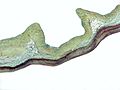 Movat's stain showing thickening of the spongiosa layer (blue) in myxomatous degeneration of the aortic valve


References
See also
Wikiwand - on
Seamless Wikipedia browsing. On steroids.
Remove ads