Top Qs
Timeline
Chat
Perspective
Neurofilament light polypeptide
Protein-coding gene in the species Homo sapiens From Wikipedia, the free encyclopedia
Remove ads
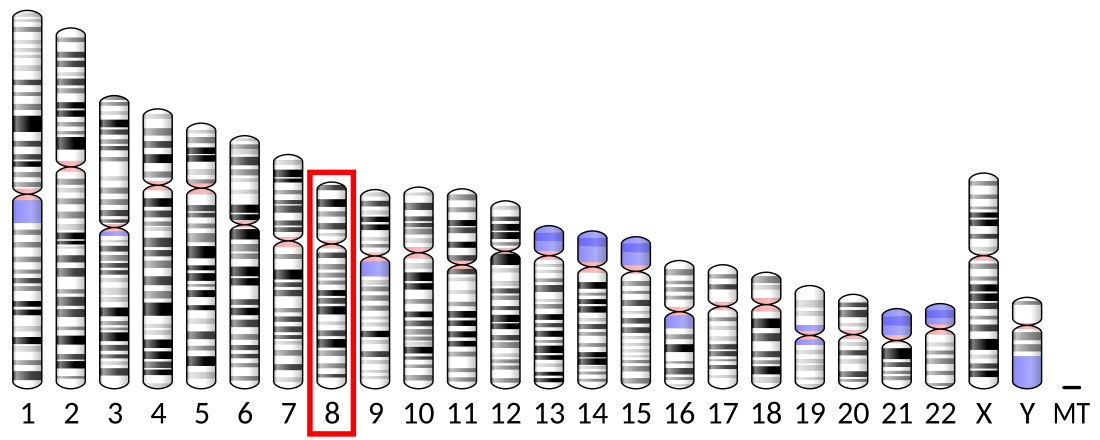
Neurofilament light polypeptide is a protein that in humans is encoded by the NEFL gene.[5]
Remove ads
Structure
Neurofilament light polypeptide is a member of the intermediate filament protein family. This protein family consists of over 50 human proteins divided into 5 major classes, the Class I and II keratins, Class III vimentin, GFAP, desmin and the others, the Class IV neurofilaments and the Class V nuclear lamins. There are four major neurofilament subunits, NF-L, NF-M, NF-H and α-internexin. These form heteropolymers which assemble to produce 10 nm neurofilaments which are only expressed in neurons where they are major structural proteins, particularly concentrated in large projection axons. The NF-L protein is encoded by the NEFL gene.[6][5]
Remove ads
Function
These neurofilament heteropolymers assemble into the cytoskeleton of axons, where they provide structural support and help regulate axonal diameter and conduction velocity. Axons are particularly sensitive to mechanical and metabolic compromise and as a result axonal degeneration is a significant problem in many neurological disorders. Neurofilament light chain is a biomarker that can be measured with immunoassays in cerebrospinal fluid and plasma and reflects axonal damage in a wide variety of neurological disorders.[7]
Remove ads
Measurement
NF-L antibodies employed in the most widely used NF-L assays are specific for cleaved forms of NF-L generated by proteolysis induced by cell death.[8] Methods used in different studies for NfL measurement are sandwich enzyme-linked immunosorbent assay (ELISA), electrochemiluminescence, and high-sensitive single molecule array (SIMOA).[9]
Clinical significance
The detection of neurofilament subunits in CSF and blood has become widely used as a biomarker of ongoing axonal compromise. It is a useful marker for disease monitoring in amyotrophic lateral sclerosis,[10] multiple sclerosis,[11] Alzheimer's disease,[12][13] and more recently Huntington's disease.[14] It is also a promising marker for follow-up of patients with brain tumors.[15] Higher levels of blood or CSF NF-L have been associated with increased mortality, as would be expected as release of this protein reflects ongoing axonal loss.[16]
It is associated with Charcot–Marie–Tooth disease 1F and 2E.[6]
Remove ads
Neurofilament assembly
Summarize
Perspective


Neurofilament light polypeptide (NF-L) is a key structural component of the neuronal cytoskeleton, assembling into neurofilaments along with other intermediate filament proteins such as NF-M, NF-H, and α-internexin.[17][18] These proteins form obligate heteropolymers that organize into 10 nm diameter filaments,[17][18] which are selectively expressed in neurons and are particularly concentrated in axons[9]. Neurofilaments provide essential structural support, help maintain axonal diameter,[19][18] and contribute to the efficient conduction of nerve impulses.[19]
The localization and organization of NF-L in neurons can be visualized using immunohistochemical techniques. In tissue culture preparations of rat brain cells, antibodies specific to NF-L label large neurons prominently in green, revealing their extensive cytoskeletal architecture.[20] In the same cultures, staining for α-internexin in red highlights surrounding neuronal stem cells, indicating the differential expression of these intermediate filament proteins during neural development and differentiation.[18]
In histological sections of human brain tissue, NF-L can also be visualized using immunostaining. For example, in formalin-fixed and paraffin-embedded sections of the human cerebellum, an antibody specific to NF-L reveals its presence throughout various neuronal compartments[7]. The brown-stained antibody binding highlights the axonal processes of basket cells, the parallel fibers of granule cells,[21][18] the perikarya of Purkinje cells,[21] and other axonal elements. Counterstaining with a blue dye allows for the visualization of cell nuclei, delineating the granular layer on the left side of the section and the molecular layer on the right.[21] These staining patterns underscore the widespread and structurally critical role of NF-L in both developing and mature neurons.[19][18]
Remove ads
Interactions
Neurofilament light polypeptide has been shown to interact with:
References
Further reading
Wikiwand - on
Seamless Wikipedia browsing. On steroids.
Remove ads


