Top Qs
Timeline
Chat
Perspective
Pudendal canal
Aspect of human anatomy From Wikipedia, the free encyclopedia

Remove ads
The pudendal canal (also called Alcock's canal) is an anatomical structure formed by the obturator fascia (fascia of the obturator internus muscle) lining the lateral wall of the ischioanal fossa. The internal pudendal artery and veins, and pudendal nerve pass through the pudendal canal, and the perineal nerve arises within it.[1]

Remove ads
Clinical significance
Pudendal nerve entrapment can occur when the pudendal nerve is compressed while it passes through the pudendal canal.[2]
History
The pudendal canal is also known as Alcock's canal, named after Benjamin Alcock.[3]
Additional images
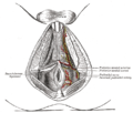 The superficial branches of the internal pudendal artery. (Canal not labeled, but pudendal nerve and internal pudendal artery labeled at bottom right.)

See also
References
External links
Wikiwand - on
Seamless Wikipedia browsing. On steroids.
Remove ads