Top Qs
Timeline
Chat
Perspective
Basilar artery
Artery that supplies the brain with blood From Wikipedia, the free encyclopedia

Remove ads
The basilar artery (U.K.: /ˈbæz.ɪ.lə/;[1][2] U.S.: /ˈbæs.ə.lər/[3]) is one of the arteries that supplies the brain with oxygen-rich blood.

The two vertebral arteries and the basilar artery are known as the vertebral basilar system, which supplies blood to the posterior part of the circle of Willis and joins with blood supplied to the anterior part of the circle of Willis from the internal carotid arteries.[4][5][6]
Remove ads
Structure
The diameter of the basilar artery range from 1.5 to 6.6 mm.[7]
Origin
The basilar artery arises from the union of the two vertebral arteries at the junction between the medulla oblongata and the pons between the abducens nerves (CN VI).[8]
Course
It ascends along the basilar sulcus of the ventral pons. It divides at the junction of the midbrain and pons into the posterior cerebral arteries.[citation needed]
Branches
Its branches from caudal to rostral include:[citation needed]
- anterior inferior cerebellar artery
- labyrinthine artery (<15% of people, usually branches from the anterior inferior cerebellar artery)
- pontine arteries
- superior cerebellar artery
Remove ads
Clinical relevance
A basilar artery stroke classically leads to locked-in syndrome.[9][10]
Additional images
 The internal carotid and vertebral arteries (Right side view)
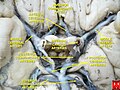 Basilar artery
 The arteries of the base of the brain. Basilar artery labeled below center. The temporal pole of the cerebrum and the cerebellar hemisphere have been removed on the right side. Inferior aspect (viewed from below).


References
External links
Wikiwand - on
Seamless Wikipedia browsing. On steroids.
Remove ads