Top Qs
Timeline
Chat
Perspective
Mesothelin
Protein found in humans From Wikipedia, the free encyclopedia
Remove ads
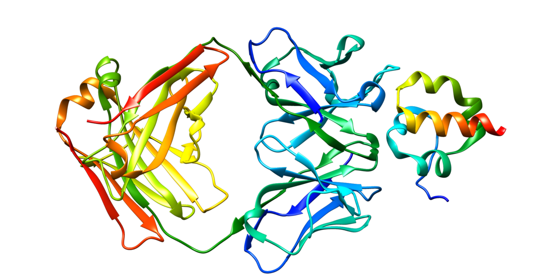
Mesothelin, also known as MSLN, is a protein that in humans is encoded by the MSLN gene.[5][6]
Remove ads
Remove ads
Function
Summarize
Perspective
Mesothelin is a 40 kDa protein that is expressed in mesothelial cells.[7] The protein was first identified by its reactivity with monoclonal antibody K1.[8] Subsequent cloning studies showed that the mesothelin gene encodes a precursor protein that is processed to yield mesothelin which is attached to the cell membrane by a glycophosphatidylinositol linkage and a 31-kDa shed fragment named megakaryocyte-potentiating factor (MPF). Although it has been proposed that mesothelin may be involved in cell adhesion, its biological function is not known.[9][10] A knockout mouse line that lacks mesothelin reproduces and develops normally.[11]
Mesothelin is over expressed in several human tumors, including mesothelioma, ovarian cancer, pancreatic adenocarcinoma,[7] lung adenocarcinoma,[12] and cholangiocarcinoma.[13] Mesothelin binds MUC16 (also known as CA125), indicating that the interaction of mesothelin and MUC16 may contribute to the implantation and peritoneal spread of tumors by cell adhesion.[14] The region (residues 296-359) consisting of 64 amino acids at the N-terminus of cell surface mesothelin has been identified as the functional binding domain (named IAB) for MUC16/CA125, suggesting the mechanism of mesothelin acting as a MUC16/CA125 functional partner in cancer development.[15]

Remove ads
Medical applications
Summarize
Perspective
Mesothelin is a tumor differentiation antigen that is normally present on the mesothelial cells lining the pleura,[17] peritoneum and pericardium.[7] Since mesothelin is overexpressed in several cancers and is immunogenic, the protein could be exploited as tumor marker or as the antigenic target of a therapeutic cancer vaccine.[9][18] A 2016 review indicates that some immunotherapeutic strategies have shown encouraging results in early-phase clinical trials.[19] Elevations of serum mesothelin specific to ovarian and other cancer patients may be measured using ELISA assays.[20] Soluble mesothelin is identified as the extracellular domain of membrane-bound mesothelin shed from tumor cells according to the mass spectrometry analysis of soluble mesothelin purified from cell culture supernatant.[21]
Assays for blood-borne mesothelin and MPF for tumor diagnosis, especially applied to asbestos-related mesothelioma have been developed.[22] Elevated serum mesothelin was found in most patients with mesothelioma (71%) and ovarian cancer (67%).[23] Blood MPF and mesothelin levels were correlated, with modest accuracy for malignant pleural mesothelioma and lung cancer (sensitivity 74% and 59%, specificity 90% and 86%, respectively for MPF and mesothelin assays).[24] Circulating mesothelin is reported in nearly all pancreatic cancers,[25] however the levels in healthy persons often exceed 80 ng/mL (using 40 kD molecular weight as the conversion factor) and to widely overlap the values in the pancreatic cancer patients.[26] It was noted that the cutoff levels for normal could differ as much as 10-fold among publications, depending on the assay used[26][24][23] and thus that normal levels must be determined anew when new assays are introduced. Increase of mesothelin-specific antibodies were also detected in the sera of about 40% of patients with mesothelioma and 42% with ovarian cancer, indicating an antibody response to mesothelin was correlated with high expression of mesothelin on tumor cells.[27]
Human monoclonal antibodies HN1 and SD1 targeting mesothelin have been isolated by phage display.[28][29] Mitchell Ho and Ira Pastan at the U.S. National Institutes of Health (NIH) generated rabbit monoclonal antibodies targeting rare and poorly immunogenic epitopes of mesothelin, including the C terminus recognized by the YP218 antibody.[16] The rabbit antibodies have been "humanized" by Ho and Zhang using human immunoglobulin germline framework sequences for CDR grafting based on computational structure modeling.[30] The CAR-T cells derived from the humanized YP218 antibody (hYP218) effectively inhibit the growth of human xenograft tumors in mice.[31][32]
Remove ads
References
Further reading
External links
Wikiwand - on
Seamless Wikipedia browsing. On steroids.
Remove ads


