Top Qs
Timeline
Chat
Perspective
Mouse brain
Body part used for neuroscience research From Wikipedia, the free encyclopedia

Remove ads
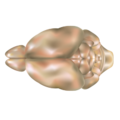
The mouse brain refers to the brain of Mus musculus. Various brain atlases for it exist.
This article's lead section may need to be rewritten. (April 2019) |
Despite superficial differences, especially in size and weight, the mouse brain and its function can serve as a powerful animal model for study of human brain diseases or mental disorders (see e.g. Reeler, Chakragati mouse). This is because the genes responsible for building and operating both mouse and human brain are 90% identical.[1] Transgenic mouse lines also allow neuroscientists to specifically target the labeling of certain cell types to probe the neural basis of fundamental processes.[2][3]
Remove ads
Anatomy
The cerebral cortex of a mouse has around 8–14 million neurons while in those humans there are more than 10–15 billion.[4][5] The olfactory bulb volume takes about 2% of the mouse brain by volume in contrast to about 0.01% of the human brain.[6][7]
 Mouse brain, dorsal view
 Mouse brain, lateral view
 Mouse brain slices
 Mouse cingulate cortex neurons
 A biophysically realistic model of a mouse's primary motor cortex microcircuit[8]


For reasons of reproducibility, genetically characterized, stable strains like C57BL/6 were chosen to produce high-resolution images and databases.[9] Well known online resources include:
Remove ads
See also
References
Wikiwand - on
Seamless Wikipedia browsing. On steroids.
Remove ads