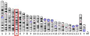
Myotilin is a protein that in humans is encoded by the MYOT gene.[5][6][7] Myotilin (myofibrillar titin-like protein) also known as TTID (TiTin Immunoglobulin Domain) is a muscle protein that is found within the Z-disc of sarcomeres.
Quick Facts Available structures, PDB ...
Close
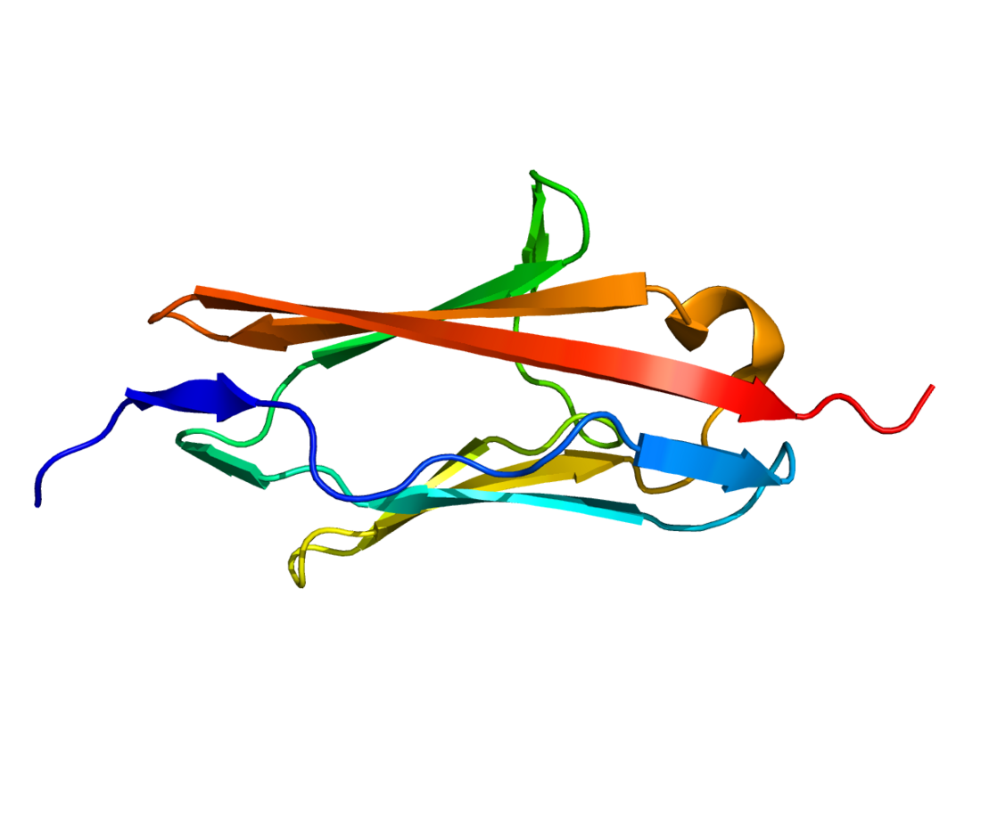
Myotilin is a 55.3 kDa protein composed of 496 amino acids.[8] Myotilin was originally identified as a novel alpha-actinin binding partner with two Ig-like domains, that localized to the Z-disc.[9] The I-type Ig-like domains reside at the C-terminal half, and are most homologous to Ig domains 2-3 of palladin and Ig domains 4-5 of myopalladin and more distantly related to Z-disc Ig domains 7 and 8 of titin. The C-terminal region hosts the binding sites for Z-band proteins, and 2 Ig domains are the site of homodimerization for myotilin.[10] By contrast, the N-terminal part of myotilin is unique, consisting of a serine-rich region with no homology to known proteins. Several disease-associated mutations involve serine residues within the serine-rich domain.[11] Myotilin expression in human tissues is mainly restricted to striated muscles and nerves. In muscles, myotilin is predominantly found within the Z-discs. Myotilin forms homodimers and binds alpha-actinin, actin,[12] Filamin C,[13] FATZ-1,[14] FATZ-2[14] and ZASP.[15]
Myotilin is a structural protein that, along with titin and alpha-actinin give structural integrity to sarcomeres at Z-discs in striated muscle. Myotilin induces the formation of actin bundles in vitro and in non-muscle cells. A ternary complex myotilin/actin/alpha-actinin can be observed in vitro and actin bundles formed under these conditions appear more tightly packed than those induced by alpha-actinin alone. It was demonstrated that myotilin stabilizes F-actin by slowing down the disassembly rate. Ectopic overexpression of truncated myotilin causes the disruption of nascent myofibrils and the co-accumulation of myotilin and titin in amorphous cytoplasmic precipitates. In mature sarcomeres, wild-type myotilin colocalizes with alpha-actinin and Z-disc titin, showing the striated pattern typical of sarcomeric proteins. Targeted disruption of the myotilin gene in mice does not cause significant alterations in muscle function.[16] On the other hand, transgenic mice with mutated myotilin develop muscle dystrophy.[17]
Myotilin is mutated in various forms of muscular dystrophy: Limb-Girdle Muscular Dystrophy type 1A (LGMD1A), Myofibrillar Myopathy (MFM), Spheroid Body Myopathy and Distal Myopath.[11] The mechanism underlying the pathology is still under investigation. It has been shown that actin binding properties of myotilin housing pathogenic mutations (Ser55Phe, Thr57Ile, Ser60Cys, and Ser95Ile) are normal,[18] albeit with a slower rate of degradation.[19] Surprisingly, YFP-fusion constructs of myotilin mutants (Ser55Phe, Ser55Ile, Thr57Ile, Ser60Cys, Ser60Phe, Ser95Ile, Arg405Lys) localized normally to Z-discs and exhibited normal dynamics in muscle cells.[20]
Godley LA, Lai F, Liu J, Zhao N, Le Beau MM (Nov 1999). "TTID: A novel gene at 5q31 encoding a protein with titin-like features". Genomics. 60 (2): 226–33. doi:10.1006/geno.1999.5912. PMID 10486214. Salmikangas P, van der Ven PF, Lalowski M, Taivainen A, Zhao F, Suila H, Schröder R, Lappalainen P, Fürst DO, Carpén O (Jan 2003). "Myotilin, the limb-girdle muscular dystrophy 1A (LGMD1A) protein, cross-links actin filaments and controls sarcomere assembly". Human Molecular Genetics. 12 (2): 189–203. doi:10.1093/hmg/ddg020. PMID 12499399. van der Ven PF, Wiesner S, Salmikangas P, Auerbach D, Himmel M, Kempa S, Hayess K, Pacholsky D, Taivainen A, Schröder R, Carpén O, Fürst DO (Oct 2000). "Indications for a novel muscular dystrophy pathway. gamma-filamin, the muscle-specific filamin isoform, interacts with myotilin". The Journal of Cell Biology. 151 (2): 235–248. doi:10.1083/jcb.151.2.235. PMC 2192634. PMID 11038172. von Nandelstadh P, Grönholm M, Moza M, Lamberg A, Savilahti H, Carpén O (Oct 2005). "Actin-organising properties of the muscular dystrophy protein myotilin". Experimental Cell Research. 310 (1): 131–9. doi:10.1016/j.yexcr.2005.06.027. PMID 16122733. von Nandelstadh P, Soliymani R, Baumann M, Carpen O (May 2011). "Analysis of myotilin turnover provides mechanistic insight into the role of myotilinopathy-causing mutations". The Biochemical Journal. 436 (1): 113–21. doi:10.1042/BJ20101672. PMID 21361873.
- Speer MC, Yamaoka LH, Gilchrist JH, Gaskell CP, Stajich JM, Vance JM, Kazantsev A, Lastra AA, Haynes CS, Beckmann JS (Jun 1992). "Confirmation of genetic heterogeneity in limb-girdle muscular dystrophy: linkage of an autosomal dominant form to chromosome 5q". American Journal of Human Genetics. 50 (6): 1211–7. PMC 1682558. PMID 1598902.
- Dixon MJ, Read AP, Donnai D, Colley A, Dixon J, Williamson R (Jul 1991). "The gene for Treacher Collins syndrome maps to the long arm of chromosome 5". American Journal of Human Genetics. 49 (1): 17–22. PMC 1683211. PMID 1676560.
- Bartoloni L, Horrigan SK, Viles KD, Gilchrist JM, Stajich JM, Vance JM, Yamaoka LH, Pericak-Vance MA, Westbrook CA, Speer MC (Dec 1998). "Use of a CEPH meiotic breakpoint panel to refine the locus of limb-girdle muscular dystrophy type 1A (LGMD1A) to a 2-Mb interval on 5q31". Genomics. 54 (2): 250–5. doi:10.1006/geno.1998.5579. PMID 9828127.
- Hauser MA, Horrigan SK, Salmikangas P, Torian UM, Viles KD, Dancel R, Tim RW, Taivainen A, Bartoloni L, Gilchrist JM, Stajich JM, Gaskell PC, Gilbert JR, Vance JM, Pericak-Vance MA, Carpen O, Westbrook CA, Speer MC (Sep 2000). "Myotilin is mutated in limb girdle muscular dystrophy 1A". Human Molecular Genetics. 9 (14): 2141–7. doi:10.1093/hmg/9.14.2141. PMID 10958653.
- van der Ven PF, Wiesner S, Salmikangas P, Auerbach D, Himmel M, Kempa S, Hayess K, Pacholsky D, Taivainen A, Schröder R, Carpén O, Fürst DO (Oct 2000). "Indications for a novel muscular dystrophy pathway. gamma-filamin, the muscle-specific filamin isoform, interacts with myotilin". The Journal of Cell Biology. 151 (2): 235–48. doi:10.1083/jcb.151.2.235. PMC 2192634. PMID 11038172.
- Hauser MA, Conde CB, Kowaljow V, Zeppa G, Taratuto AL, Torian UM, Vance J, Pericak-Vance MA, Speer MC, Rosa AL (Dec 2002). "myotilin Mutation found in second pedigree with LGMD1A". American Journal of Human Genetics. 71 (6): 1428–32. doi:10.1086/344532. PMC 378586. PMID 12428213.
- Salmikangas P, van der Ven PF, Lalowski M, Taivainen A, Zhao F, Suila H, Schröder R, Lappalainen P, Fürst DO, Carpén O (Jan 2003). "Myotilin, the limb-girdle muscular dystrophy 1A (LGMD1A) protein, cross-links actin filaments and controls sarcomere assembly". Human Molecular Genetics. 12 (2): 189–203. doi:10.1093/hmg/ddg020. PMID 12499399.
- Battle MA, Maher VM, McCormick JJ (Jun 2003). "ST7 is a novel low-density lipoprotein receptor-related protein (LRP) with a cytoplasmic tail that interacts with proteins related to signal transduction pathways". Biochemistry. 42 (24): 7270–82. doi:10.1021/bi034081y. PMID 12809483.
- Selcen D, Engel AG (Apr 2004). "Mutations in myotilin cause myofibrillar myopathy". Neurology. 62 (8): 1363–71. doi:10.1212/01.wnl.0000123576.74801.75. PMID 15111675. S2CID 23068161.
- Witt SH, Granzier H, Witt CC, Labeit S (Jul 2005). "MURF-1 and MURF-2 target a specific subset of myofibrillar proteins redundantly: towards understanding MURF-dependent muscle ubiquitination". Journal of Molecular Biology. 350 (4): 713–22. doi:10.1016/j.jmb.2005.05.021. PMID 15967462.
- Gontier Y, Taivainen A, Fontao L, Sonnenberg A, van der Flier A, Carpen O, Faulkner G, Borradori L (Aug 2005). "The Z-disc proteins myotilin and FATZ-1 interact with each other and are connected to the sarcolemma via muscle-specific filamins". Journal of Cell Science. 118 (Pt 16): 3739–49. doi:10.1242/jcs.02484. PMID 16076904.
- von Nandelstadh P, Grönholm M, Moza M, Lamberg A, Savilahti H, Carpén O (Oct 2005). "Actin-organising properties of the muscular dystrophy protein myotilin". Experimental Cell Research. 310 (1): 131–9. doi:10.1016/j.yexcr.2005.06.027. PMID 16122733.
- Rual JF, Venkatesan K, Hao T, Hirozane-Kishikawa T, Dricot A, Li N, Berriz GF, Gibbons FD, Dreze M, Ayivi-Guedehoussou N, Klitgord N, Simon C, Boxem M, Milstein S, Rosenberg J, Goldberg DS, Zhang LV, Wong SL, Franklin G, Li S, Albala JS, Lim J, Fraughton C, Llamosas E, Cevik S, Bex C, Lamesch P, Sikorski RS, Vandenhaute J, Zoghbi HY, Smolyar A, Bosak S, Sequerra R, Doucette-Stamm L, Cusick ME, Hill DE, Roth FP, Vidal M (Oct 2005). "Towards a proteome-scale map of the human protein-protein interaction network". Nature. 437 (7062): 1173–8. Bibcode:2005Natur.437.1173R. doi:10.1038/nature04209. PMID 16189514. S2CID 4427026.
- Foroud T, Pankratz N, Batchman AP, Pauciulo MW, Vidal R, Miravalle L, Goebel HH, Cushman LJ, Azzarelli B, Horak H, Farlow M, Nichols WC (Dec 2005). "A mutation in myotilin causes spheroid body myopathy". Neurology. 65 (12): 1936–40. doi:10.1212/01.wnl.0000188872.28149.9a. PMID 16380616. S2CID 9593230.
- Garvey SM, Senderek J, Beckmann JS, Seboun E, Jackson CE, Hauser MA (May 2006). "Myotilin is not the causative gene for vocal cord and pharyngeal weakness with distal myopathy (VCPDM)". Annals of Human Genetics. 70 (Pt 3): 414–6. doi:10.1111/j.1529-8817.2005.00252.x. PMID 16674563. S2CID 26853063.
- Pénisson-Besnier I, Talvinen K, Dumez C, Vihola A, Dubas F, Fardeau M, Hackman P, Carpen O, Udd B (Jul 2006). "Myotilinopathy in a family with late onset myopathy". Neuromuscular Disorders. 16 (7): 427–31. doi:10.1016/j.nmd.2006.04.009. PMID 16793270. S2CID 21589529.
- Zong NC, Li H, Li H, Lam MP, Jimenez RC, Kim CS, Deng N, Kim AK, Choi JH, Zelaya I, Liem D, Meyer D, Odeberg J, Fang C, Lu HJ, Xu T, Weiss J, Duan H, Uhlen M, Yates JR, Apweiler R, Ge J, Hermjakob H, Ping P (Oct 2013). "Integration of cardiac proteome biology and medicine by a specialized knowledgebase". Circ Res. 113 (9): 1043–53. doi:10.1161/CIRCRESAHA.113.301151. PMC 4076475. PMID 23965338.
- GeneReviews/NIH/NCBI/UW entry on Myofibrillar Myopathy
- Overview of all the structural information available in the PDB for UniProt: Q9UBF9 (Myotilin) at the PDBe-KB.


